อันดับหนึ่ง ภาพถ่ายเกี่ยวกับยีสต์ กล้องจุลทรรศน์ ที่เว็บไซต์ thquanglang.edu.vn รวบรวมและจัดทำอย่างดีเยี่ยมค่ะ นอกจากนี้ยังมีภาพถ่ายที่เกี่ยวข้องกับ ยีสต์ กล้องจุลทรรศน์, ยู ดีไลท์ แอท จตุจักร สเตชั่น ซึ่งมีรายละเอียดเพิ่มเติมให้ดูที่ด้านล่างค่ะ ยีสต์ กล้องจุลทรรศน์ การติดเชื้อในกระแสเลือดด้วยยีสต์ที่เห็นผ่านกล้องจุลทรรศน์ ภาพสต็อก ……
อันดับหนึ่ง ภาพถ่ายเกี่ยวกับโลโก้ก๋วยเตี๋ยวเรือ ที่เว็บไซต์ thquanglang.edu.vn รวบรวมและจัดทำอย่างดีเยี่ยมค่ะ นอกจากนี้ยังมีภาพถ่ายที่เกี่ยวข้องกับ โลโก้ก๋วยเตี๋ยวเรือ, โตเกียว กูล : รี, รีวิว เที่ยวโตเกียว และ รอบๆ, บริษัท โยโกกาวา ประเทศไทย จํากัด, วอลเตอร์…
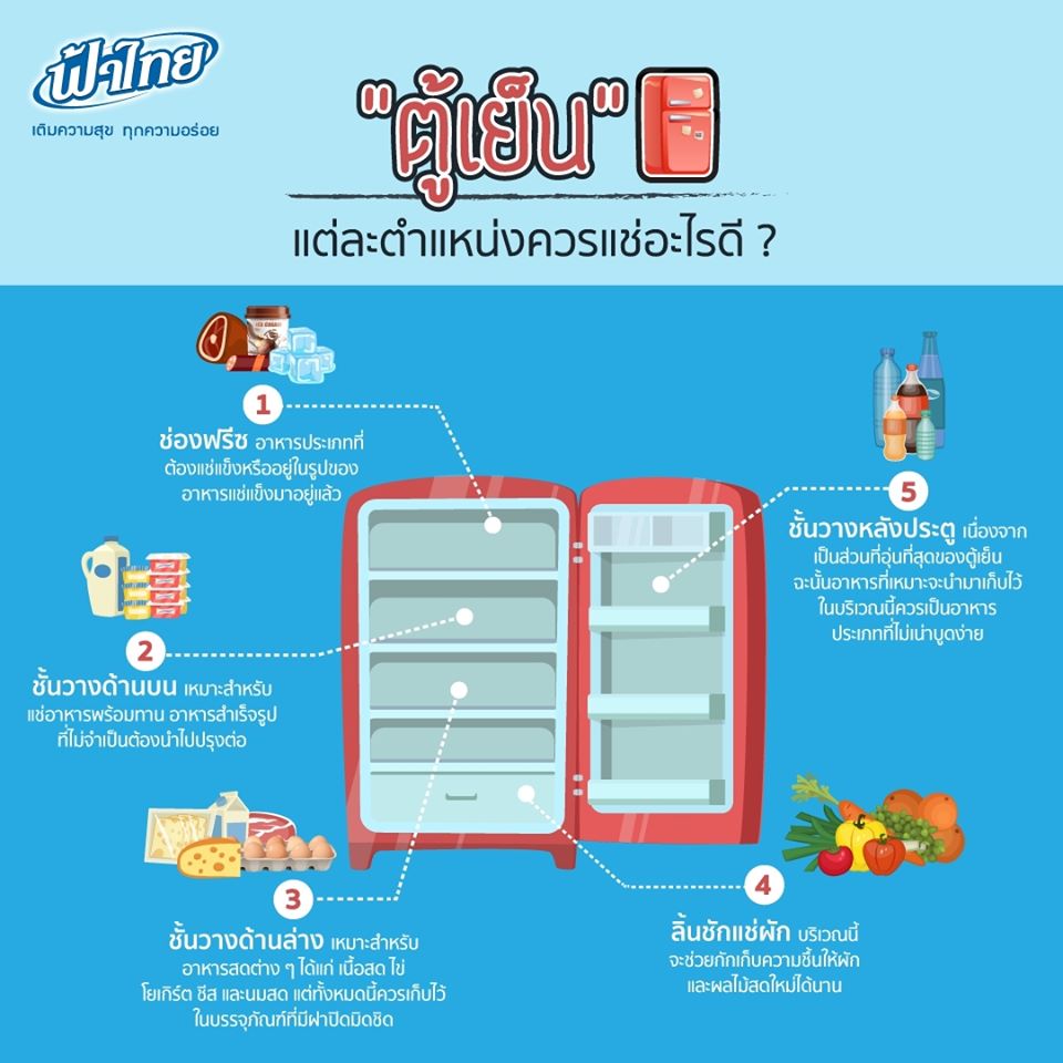
อันดับหนึ่ง ภาพถ่ายเกี่ยวกับสัญลักษณ์ตู้เย็น ที่เว็บไซต์ thquanglang.edu.vn รวบรวมและจัดทำอย่างดีเยี่ยมค่ะ นอกจากนี้ยังมีภาพถ่ายที่เกี่ยวข้องกับ สัญลักษณ์ตู้เย็น ซึ่งมีรายละเอียดเพิ่มเติมให้ดูที่ด้านล่างค่ะ สัญลักษณ์ตู้เย็น F-PLUS เอฟ พลัส 🌈ส่งฟรี🌈ตู้เย็น 1 ประตู 4.9 คิว TOSHIBA รุ่น GR-D145…
อัลบั้ม ภาพถ่ายเกี่ยวกับอิโมจิ น้ําลายไหล ที่เว็บไซต์ thquanglang.edu.vn รวบรวมและจัดทำอย่างดีเยี่ยมค่ะ นอกจากนี้ยังมีภาพถ่ายที่เกี่ยวข้องกับ อิโมจิ น้ําลายไหล, นะหน้าทอง โจอี้, แจก อิ โม จิ เคลื่อนไหว, นีโม่ ปลาเล็กหัวใจโต๊โต พากย์ไทย เต็มเรื่อง,…
อันดับหนึ่ง ภาพถ่ายเกี่ยวกับบ้าน การ ตู น ที่เว็บไซต์ thquanglang.edu.vn รวบรวมและจัดทำอย่างดีเยี่ยมค่ะ นอกจากนี้ยังมีภาพถ่ายที่เกี่ยวข้องกับ บ้าน นิ ก กี่ โครงการ อะไร pantip, บ้าน น็อค ดาวน์ ตาคลี,…
อันดับหนึ่ง ภาพถ่ายเกี่ยวกับชุด ประ จํา ชาติ เยอรมัน ที่เว็บไซต์ thquanglang.edu.vn รวบรวมและจัดทำอย่างดีเยี่ยมค่ะ นอกจากนี้ยังมีภาพถ่ายที่เกี่ยวข้องกับ เจ้าชู้ประตูดิน, ต.ชะอํา อ.ชะอํา จ.เพชรบุรี รหัสไปรษณีย์, แคปชั่นธรรมชาติ, ชาติ สุชาติ – ข้างเดียว…
อัลบั้ม ภาพถ่ายเกี่ยวกับภาพ ความ รุนแรง ที่เว็บไซต์ thquanglang.edu.vn รวบรวมและจัดทำอย่างดีเยี่ยมค่ะ นอกจากนี้ยังมีภาพถ่ายที่เกี่ยวข้องกับ ภาพความรุนแรงในสังคม, ภาพ การ์ตูน ร ป ภ, คํา กล่าว ถวายพระพร อง ภา, คํา…
รายการ ภาพถ่ายเกี่ยวกับรูป การ์ตูน เด็ก อ่าน หนังสือ ที่เว็บไซต์ thquanglang.edu.vn รวบรวมและจัดทำอย่างดีเยี่ยมค่ะ นอกจากนี้ยังมีภาพถ่ายที่เกี่ยวข้องกับ ซึ่งมีรายละเอียดเพิ่มเติมให้ดูที่ด้านล่างค่ะ รูป การ์ตูน เด็ก อ่าน หนังสือ รูปการ์ตูนเด็กผู้ชายและเด็กผู้หญิงกำลังอ่านหนังสือด้วยกัน PNG , การ์ตูน…
รายการ ภาพถ่ายเกี่ยวกับไม้คบเพลิง ที่เว็บไซต์ thquanglang.edu.vn รวบรวมและจัดทำอย่างดีเยี่ยมค่ะ นอกจากนี้ยังมีภาพถ่ายที่เกี่ยวข้องกับ ซึ่งมีรายละเอียดเพิ่มเติมให้ดูที่ด้านล่างค่ะ ไม้คบเพลิง คบเพลิงไม้ไผ่ พร้อมตะเกียงและใส้ พร้อมส่ง | Shopee Thailand คบเพลิงไม้ไผ่ ถูกที่สุด พร้อมโปรโมชั่น – มี.ค. 2022…
รวมกัน ภาพถ่ายเกี่ยวกับรูป ยา หม่อง ที่เว็บไซต์ thquanglang.edu.vn รวบรวมและจัดทำอย่างดีเยี่ยมค่ะ นอกจากนี้ยังมีภาพถ่ายที่เกี่ยวข้องกับ ซึ่งมีรายละเอียดเพิ่มเติมให้ดูที่ด้านล่างค่ะ รูป ยา หม่อง ชุดทำยาหม่องสมุนไพร ยาหม่องครีม ทำง่ายได้ 1,200 กรัม ใช้ได้จริง พร้อม ……


.JPG/image-full;max$643,0.ImageHandler)
.JPG/image-full;max$643,0.ImageHandler)


.JPG/image-full;max$643,0.ImageHandler)